Neuron Up Close and Personal
Images were taken with a scanning electron microscope, digitized, and then animated. Created by Tina (Weatherby) Carvalho.
Go see this interesting animation of a real neuron
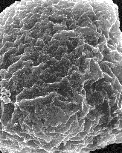
The next image is also from Microangela it is a single neuron which has been grown in a petri dish. The lump at the far end is the cell body. The flat part spread out at the closer end is the growth cone area, a part of the neuron that is trying to find the cell(s) it’s supposed to make a synapse with. The skinny part in between is the axon. The little hairs sticking up from the cell body are possibly forming dendrites.

MicroAngela is a website with a lot of Electron Microscope Images. Images may be downloaded from the site for educational, non-profit use only (such as a school report). If you need higher resolution images, without the watermark, you can contact Tina Carvalho. Terrific, spread the word…….
 GoodReads
 Last
Last
Weekly Noggin Raisers « N e u r o n a r r a t i v e
December 12, 2008 @ 10:27 pm
[…] Dr. Shock gets us up close and personal with a neuron – great animated view you won’t see very often […]
Points of Interest, #38 « Mind, Soul, and Body
December 13, 2008 @ 7:48 am
[…] Shock, MD has a fantastic electron microscope slide show that takes you up close and personal with the neuron, the cellular base of all thought, sensation, and action, turns out it looks cool, […]
December 26, 2008 @ 9:56 pm
Dr. Shock,
Very cool images. I’ve seen lots of drawings of neurons and axons, and such, as a multiple sclerosis patient. But I’d had yet to see an actual image. Thanks.